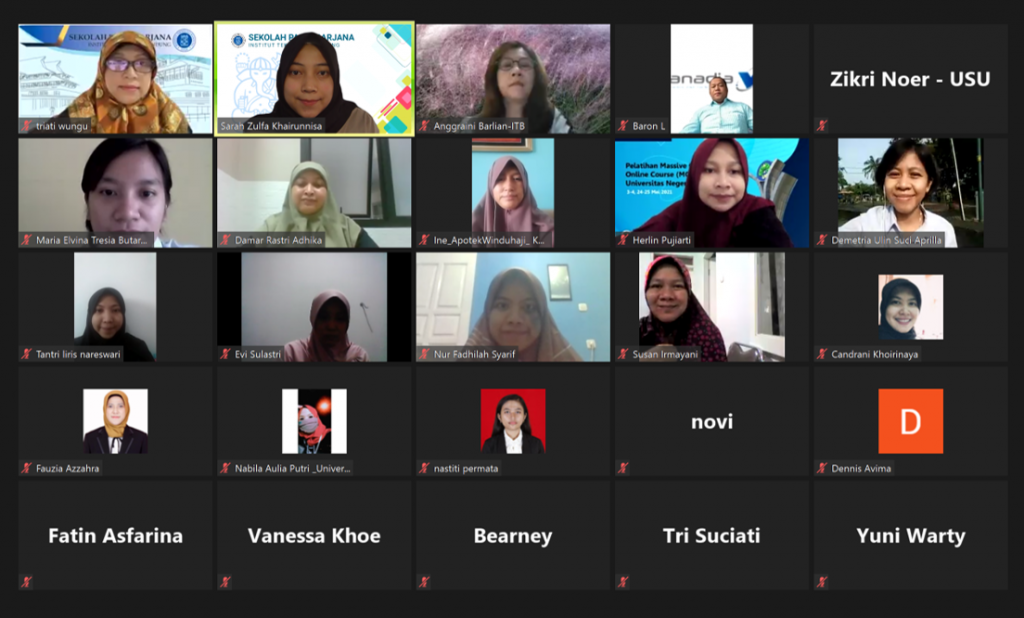
The activity will be held on Wednesday 5 May 2021, via a Webinar with the Zoom Platform. The aim of this workshop is to introduce techniques for observing biological samples using a confocal microscope. Material will be given regarding the basic concept of confocal and its application in research in the field of biological science and technology, especially related to stem-cell. The confocal instrument from Carl Zeiss LSM 900 which uses the latest technology (Airyscan.2) will be introduced by PT Vanadia Utama. Participants will be given a certificate for their participation in this webinar. To see all the activities of this webinar, click here.
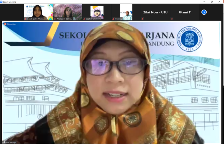
The event started with opening and remarks from Dr. triati . Then proceed with material from Dr. Damar Rastri Adhika regarding Microscopy in Life Science.
Dr. Damar Rastri conveyed that the types of microscopy that can be used in live science applications include optical microscopes, electron microscopes, and Scanning Probe Microscope(SPM). The choice of using a microscope can be adjusted to the advantages and disadvantages of each system. In general, higher resolution can be obtained by using an electron microscope which has a smaller wavelength, by calculations according to Abbe Theory. The SPM itself can be used by considering the condition of the sample due to the interaction between the probe used and the sample being observed.

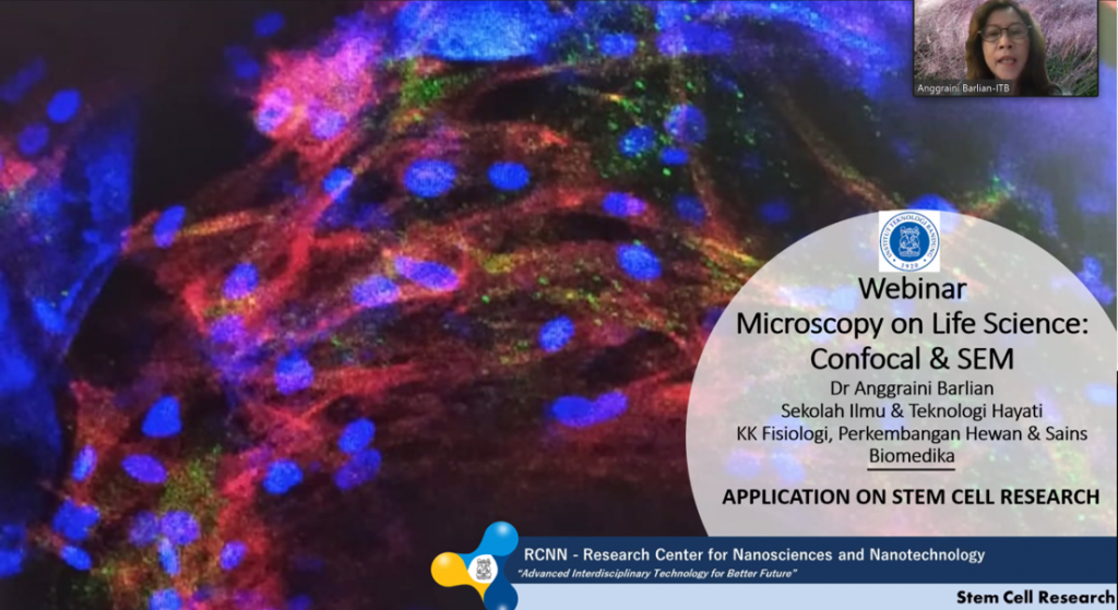
The material was continued by Dr. Anggraini Barlian, M.Sc. regarding Application on Stem Cell Research. Dr. Anggraini said that the role of the microscope in Life Scienceis very large. Research using stem cells is very dependent on the microscope because of the changes that occur inside the cell as well as outside the cell. During the differentiation process it can be visualized and confirmed by microscopy. Cell structure can be observed by SEM (Scanning Electron Microscopy) and TEM (Transmission Electron Microscopy). In addition, cytoskeletal components and the location of marker molecules can be observed using a confocal microscope.
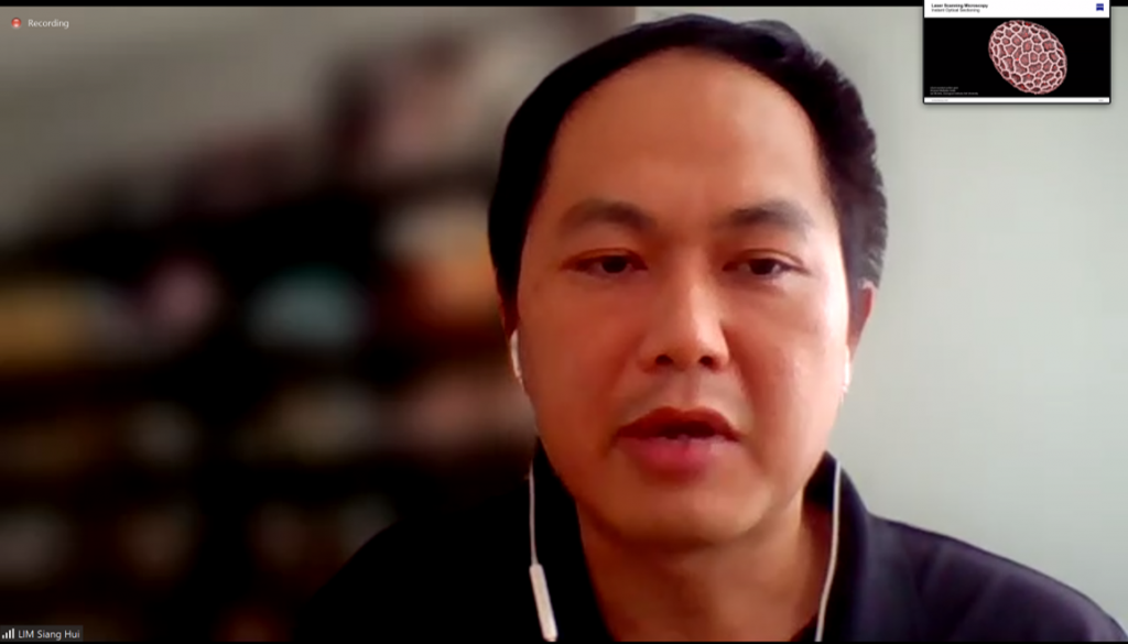
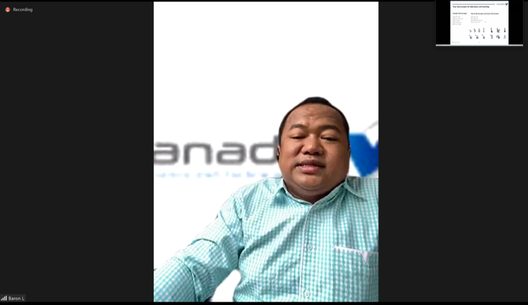
Then the material was continued by Dr. Shang Hui regarding Confocal Application and material from Baron Leonard, ST regarding Product Presentation.